Technical Images
Paintings can be imaged at various wavelengths on the electromagnetic spectrum. In addition to the visible light range, infrared, x-ray and ultraviolet frequencies reveal additional information.


Infrared Refloctography
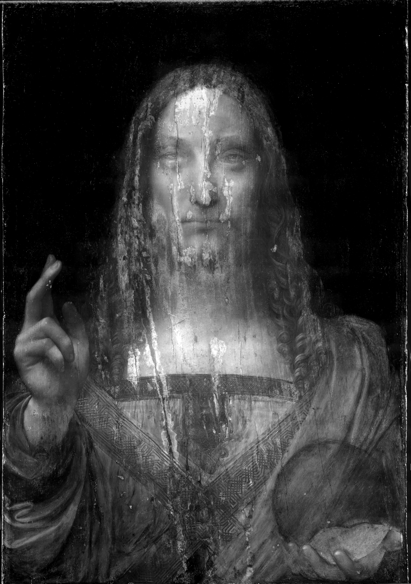
The study of paintings with infrared wavelengths has a long history. Van Asperen de Boer first used infrared film at approximately 900 nm. 11 Infrared reflectography records a wider range of wavelengths and penetrates more deeply into the paint layers. Carbon black easily absorbs infrared light and the technique is therefore very effective for obtaining images of underdrawing or underpainting when these preparatory stages were executed on a white ground with pigments containing carbon black.
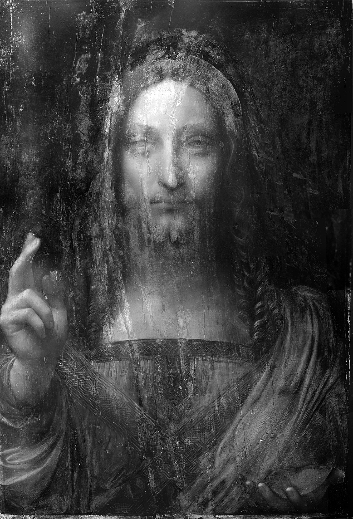
IRR scan. September 2007. Composite of captures made with a video camera fitted with a Hamamatsu vidicon with a .9 - 1.6 nm range
___________________________________ courtesy of Salvator Mundi, LLC
___________________________________ courtesy of Salvator Mundi, LLC
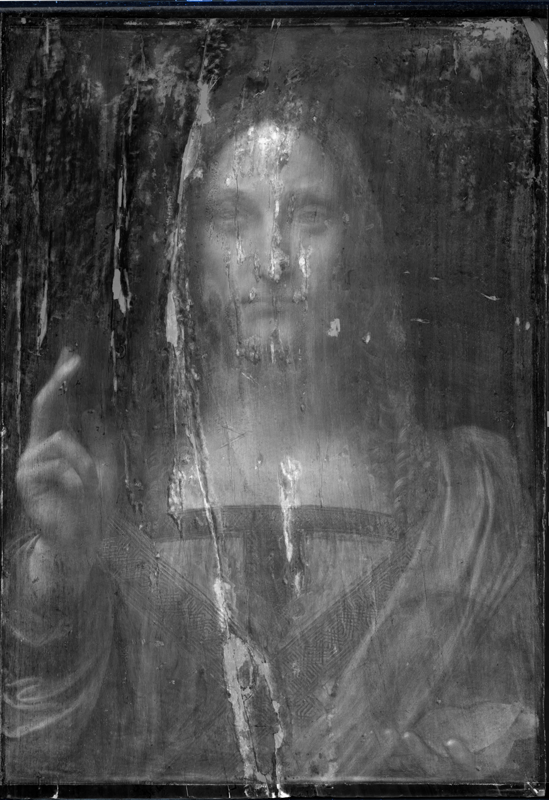
Infrared Scan of Head
Indigo Systems Merlin InGaAs near infrared digital camera sensitive to wavelengths from 900 to 1700 nm
IRR by Charlotte Hale, MMA, Courtesy of Salvator Mundi, LLC
Indigo Systems Merlin InGaAs near infrared digital camera sensitive to wavelengths from 900 to 1700 nm
IRR by Charlotte Hale, MMA, Courtesy of Salvator Mundi, LLC
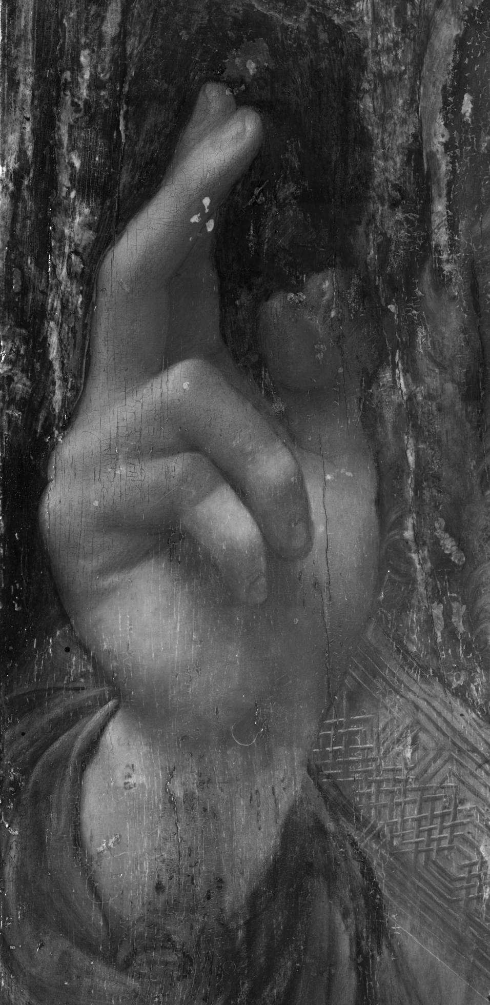
Infrared scan of blessing hand
IRR by Charlotte Hale, MMA, Courtesy of Salvator Mundi, LLC
IRR by Charlotte Hale, MMA, Courtesy of Salvator Mundi, LLC
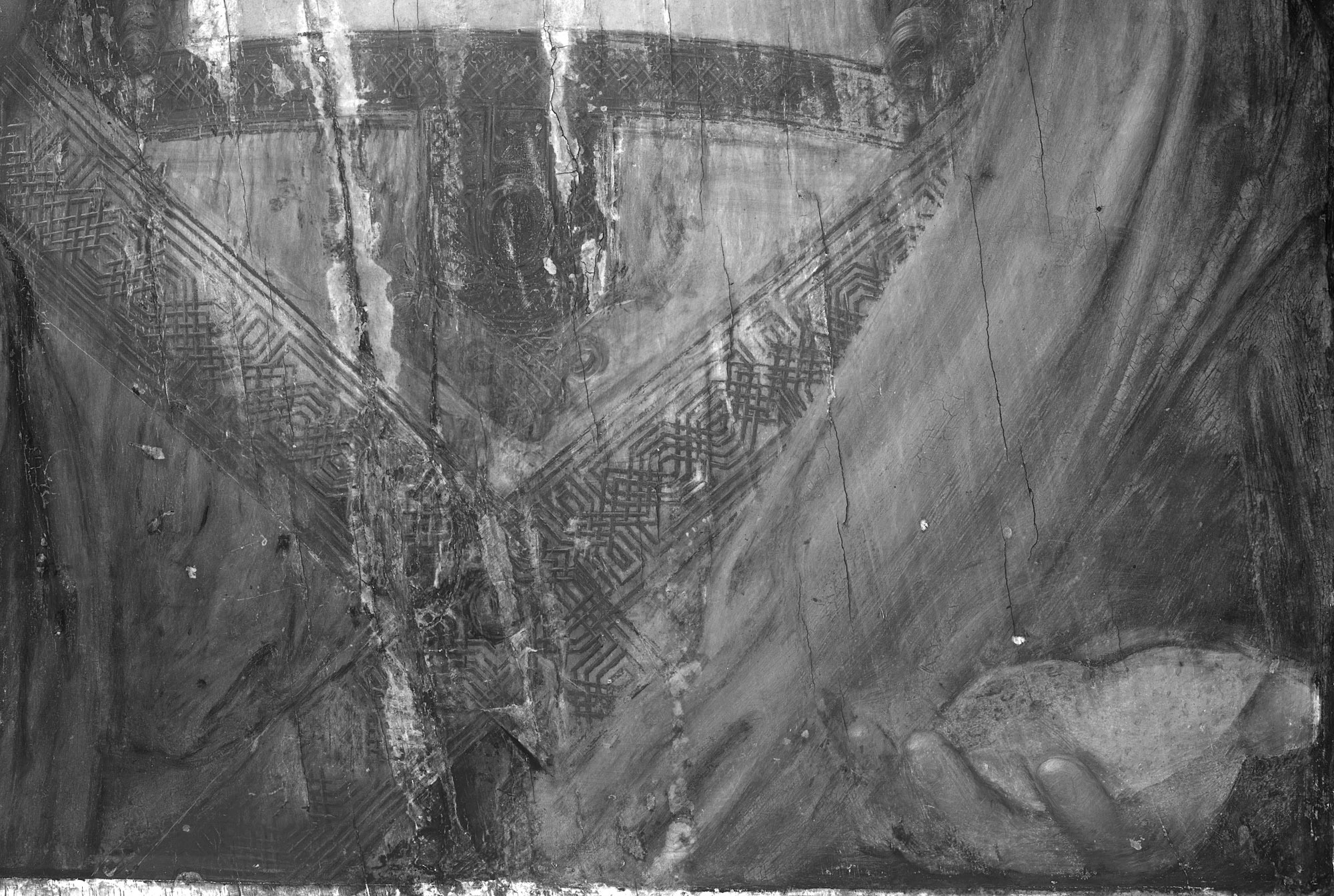
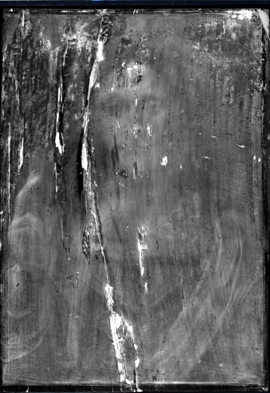
Infrared scan of the drapery with stole and orb
IRR by Charlotte Hale, MMA, Courtesy of Salvator Mundi, LLC
IRR by Charlotte Hale, MMA, Courtesy of Salvator Mundi, LLC
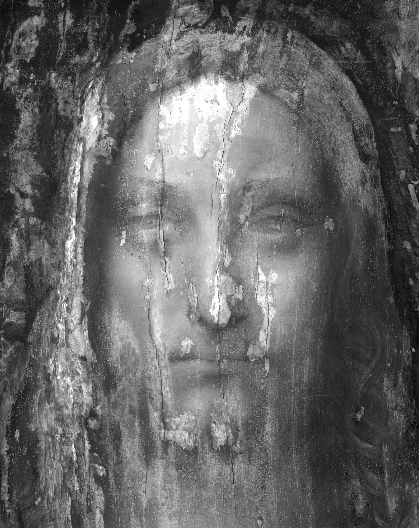
Infrared scan
FLIR SC2500-NIR InGaAs camera with 0.9 - 1.7 nm range and IR Vista software
by Shan KuanG, Conservation Center
FLIR SC2500-NIR InGaAs camera with 0.9 - 1.7 nm range and IR Vista software
by Shan KuanG, Conservation Center
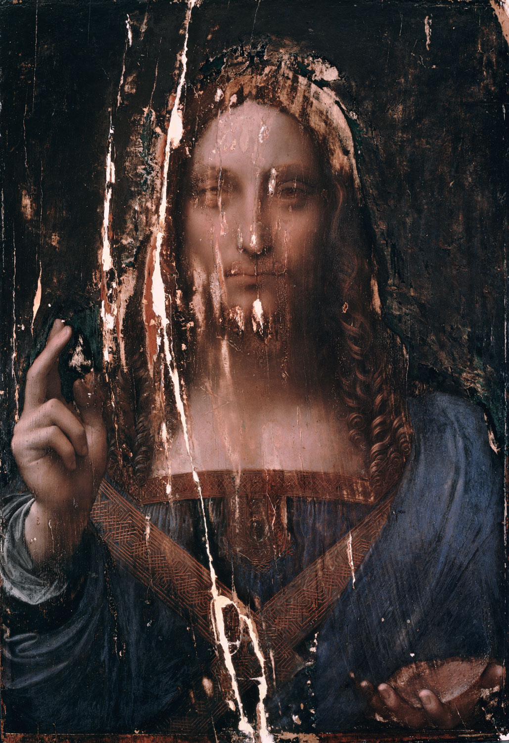
Digital X-radiograph
X-rays penetrate all the materials of a painting, including the support and are absorbed by layers containing such heavy elements as lead. The resulting image essentially records the distribution of these elements, primarily contained by the pigments lead white and lead tin yellow that were used in the preparatory layers and the paint itself.
X-radiographs of paintings by Leonardo typically exhibit a spectral appearance due to the lead white preparation, the fluidity of medium, and the many layered build-up of the sfumato. A good comparison is the X-radiograph of Saint John.
Digital X-radiographCarestream HPx-1 digital system
by Shan KuanG, Conservation Center
by Shan KuanG, Conservation Center
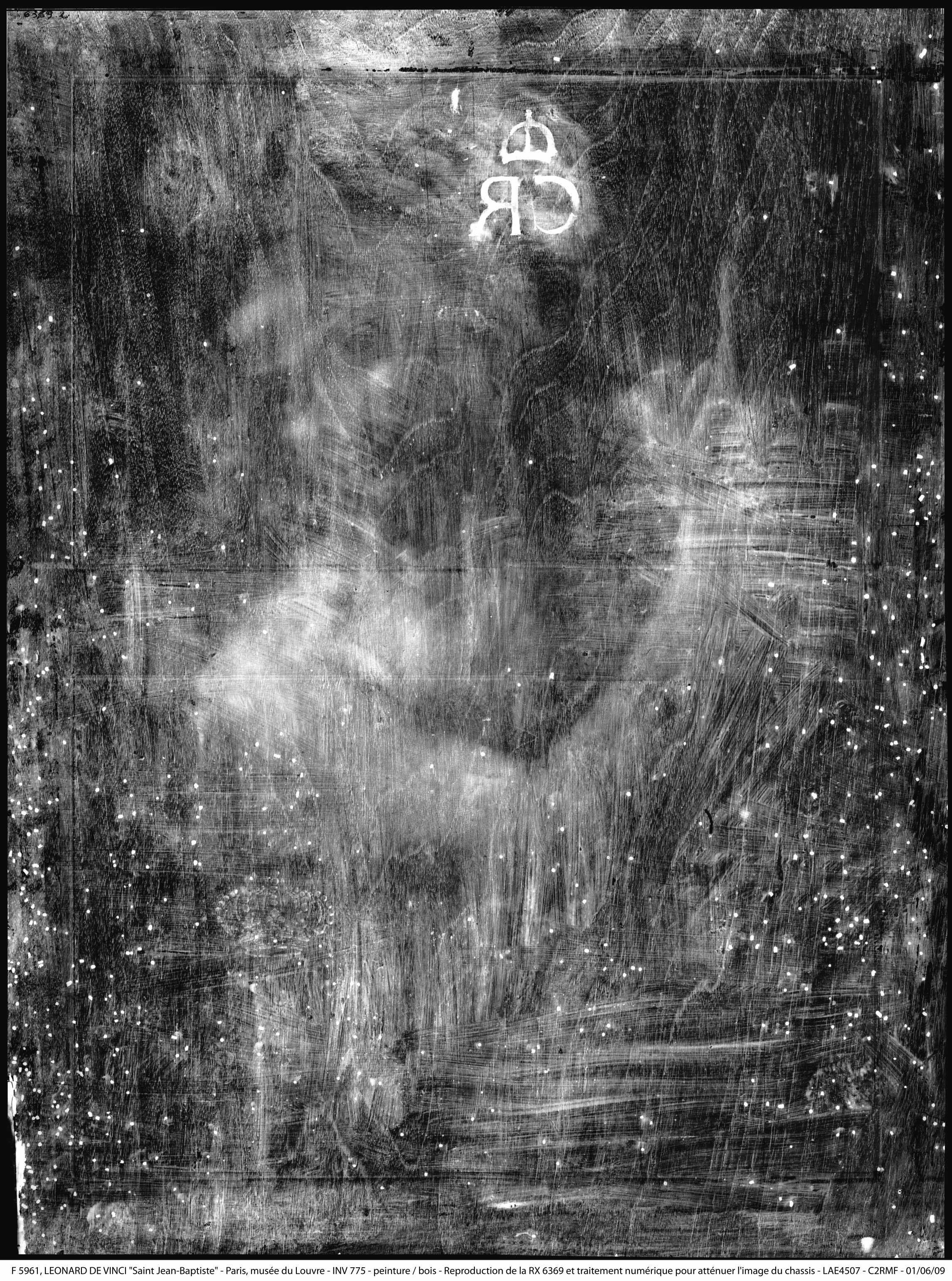
X-radiograph of Saint John
Scala/art resource
Scala/art resource
Ultraviolet Photograph
Long wave UV examination reveals the presence of varnish, which fluoresces and appears as a yellow green haze over the surface. The drying oils of the binder for the original paint also fluoresce and some pigments have a natural fluorescence of their own such as lead white and certain lake pigments, particularly madder lake. Areas of retouching exhibit no fluorescence but reflect back the dark color of the lamp and appear dark or black.
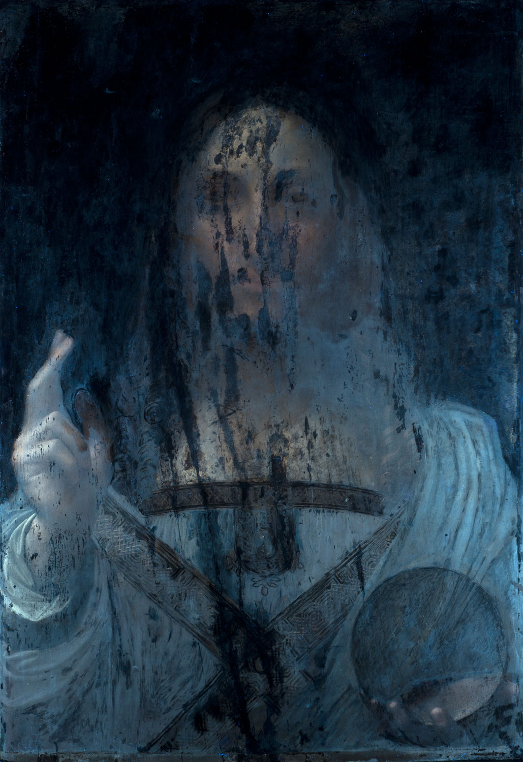
Ultraviolet Photograph
by Shan KuanG, Conservation Center
by Shan KuanG, Conservation Center