Scientific analysis
Nine samples were taken from the painting by Nica Gutman Rieppi. They were embedded in Bioplast® resin, cured, and polished as cross-sections. Ms. Rieppi sought assistance from two scientists at the Philadelphia museum of art, Dr. Beth Price and Dr. Kenneth Sutherland, who carried out scientific analyses on these cross-sections. At a later date another sample was taken for media analysis. Dr. Price and Dr. Sutherland enlisted several of their colleagues for specialized procedures: Dr. Thomas J. Tague jr. of Bruker Biospin Corporation, Billerica, MA, and Dr. Richard Newman of the Museum of Fine Arts, Boston. Ms. Rieppi coordinated the research and made notes, from which she prepared a preliminary report as the work progressed. The information in this section is drawn from that report. The scientists will eventually publish the results of their findings in a peer-reviewed journal.
Pigment Identification
The cross-sections were examined in visible and ultraviolet (UV) light with a Leitz Laborlux S microscope at the Philadelphia Museum of Art. A Leitz D filter cube was used (355-425 nm excitation, 460 nm suppression filter), and a 100 W mercury source. Fiber optic lights were used for visible light. Samples were photographed using a Nikon Digital Sight DS-5M camera with Nikon Eclipse Net image capture software.
In addition to polarized light microscopy, the pigments were identified by further analyses including x-ray fluorescence (XRF); Raman spectroscopy; Fourier transform infrared micro spectroscopy (MFTIR); and attenuated total reflection (ATR-FTIR). Scanning electron microscopy with energy dispersive spectroscopy (SEM-EDS) identified the blue mineral lazurite.
The following pigments were found in the original paint layers:
- White lead
- Iron oxide (hematite)
- Vermilion
- Bone black
- Carbon black
- Lapis Lazuli (lazurite)
- Lead tin yellow
- Red Lake (possibly madder, judging from the fluoresence)
From Nica Gutman Rieppi’s notes:
Soda lime glass particles of varying sizes were identified in the preparatory layers and in some of the paint layers, particularly as an addition to the vermilion and carbon black of the reddish brown translucent underpaint.
For Raman analyses, samples were examined as paint cross-sections using a Bruker Senterra Raman microscope equipped with a 785 nm laser and 50x objective. Data were collected using ~ 3 cm1 resolution between 70 and 3,500 cnr1 with a 20 second integration time and 2 accumulations per spectrum. The power on the samples was ~1 m to prevent transient heating. Area Raman images also were collected using an Infinity 1 digital video camera and relevant spectra extracted from the video images. All data were processed using Bruker OPUS 6.5 software. Fluorescence and polarized light illumination facilitated sample observation and characterization. Analyses were performed with the assistance of Peng Wang and Ting Wang, Applications Scientists, at Bruker Inc., Billerica, MA.
For SEM-EDS analyses, paint cross-sections were mounted on carbon tape on an aluminum stub. One cross-section was carbon-coated using a Denton Desk II cold sputter coater with carbon rod evaporation accessory. Backscattered electron imaging and EDS analyses were performed using a JEOL 6460LV SEM with an Oxford Instruments INCA X-sight EDS detector and INCA Energy 200 software. An accelerating voltage of 20 kV was used. The carbon coated cross-section was run under high vacuum and the remaining cross-sections under low vacuum at 30 Pascal.
For MFTIR analysis, the preparatory layers were isolated and representative portions flattened on a Spectra-Tech diamond window with a roller tool for analysis. Analyses were undertaken at the Philadelphia Museum of Art using a Thermo Nicolet Continuum microscope (MCT-A detector) attached to a Nexus 670 spectrometer. The data were collected in transmission mode between 4000 and 600 cm4 at 4 cnr1 resolution and 200 scans per spectrum. Omnic 6.0 software was used; spectra were processed using Happ-Genzel apodization.
Lead white pigment, consisting of both neutral and basic lead carbonate, was identified by MFTIR and EDS. Diagnostic IR spectral bands included very strong carbonate stretches centered at 1421 cm1 with additional bands at 1050, 883 and 679 cnr1 for the neutral form and at 1045 and 650 cm1 for the basic form. Lead (Pb) was detected by EDS. Lead tin yellow type I pigment (PbzSnCU) was indicated by characteristic Raman spectral bands at 127,129, 196, 273, 292, 456, and 526 cm1 and the detection of lead (Pb) and tin (Sn) by EDS.IR spectral bands suggesting an oil-based medium were observed at 2929/2851,1739 and 1170 cm1.
Iron oxide was indicated by the detection of iron (Fe) using EDS. Additional analysis by Raman produced spectra consistent with hematite, with bands located at 224, 292, and 409 cm1. Weak bands also were observed at 495 and 610 cm1.
Lazurite (lapis lazuli) was identified with diagnostic Raman spectral bands were located at 1096 and 548 cm-1 and IR spectral bands at 1114 (994 shoulder) and 2340 cm1. The IR band at 2340 cm1 is considered distinctive and thought to derive only from lapis lazuli from Afghanistan. It has not been reported in spectra of lapis lazuli from other geographical regions or of synthetic ultramarine. No copper (Cu) was detected in the cross-sections by EDS to suggest azurite [Cu3(C03)2(OH)2] nor was this pigment detected by MFTIR or Raman.
Carbon black was indicated by Raman spectra exhibiting bands at 1580 and 1325 cm1.
Bone black was indicated at 961 cm1. The identification of bone black [mainly Ca(P0)4(0H)] was supported by the detection of both calcium (Ca) and phosphorous (P) by EDS.
Vermilion: Mercury (Hg) and sulfur (S) indicative of vermilion (HgS) were detected by EDS. Raman analysis produced spectra with characteristic bands for vermilion at 255 and 344 cm1.
Red Lake: Some pigment particles fluoresced an orange-pink color when excited with UV-violet (355-425 nm) light using FLM. Aluminum (Al), suggesting an alum derived substrate, was detected by EDS. The red colorant was not present in sufficient quantities to enable analysis by such supplemental techniques as high performance liquid chromatography or mass spectrometry.
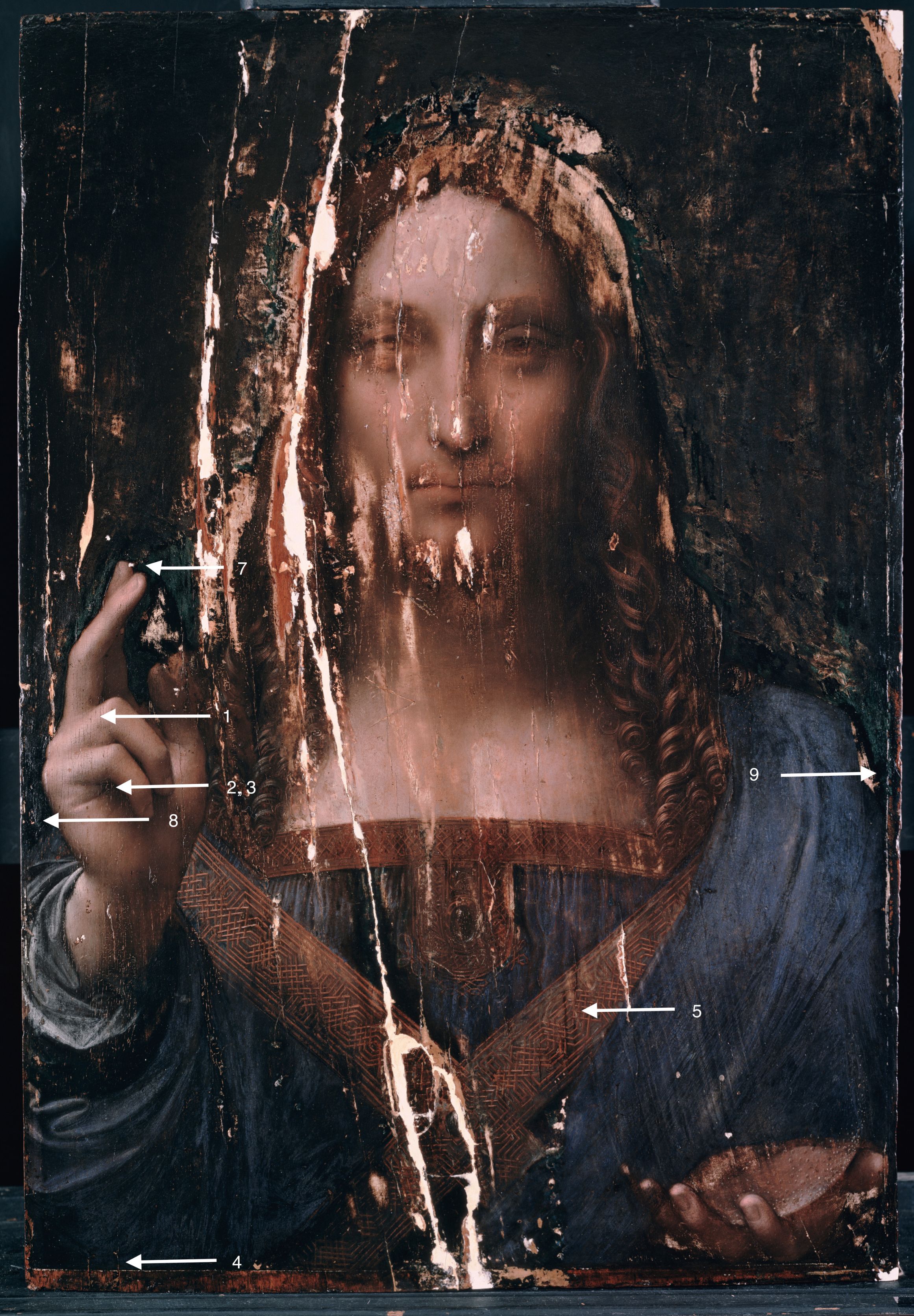
Locations of the 9 samples
Cross Sections
All images were made by Nica Gutman Rieppi. I have edited her original notations for consistency.
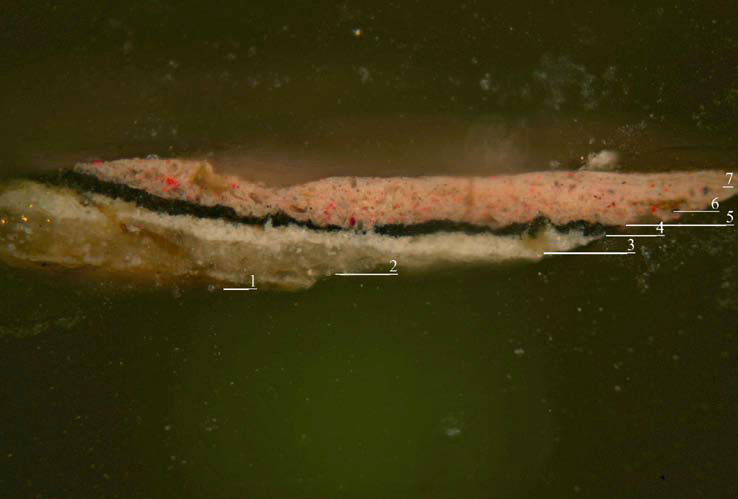
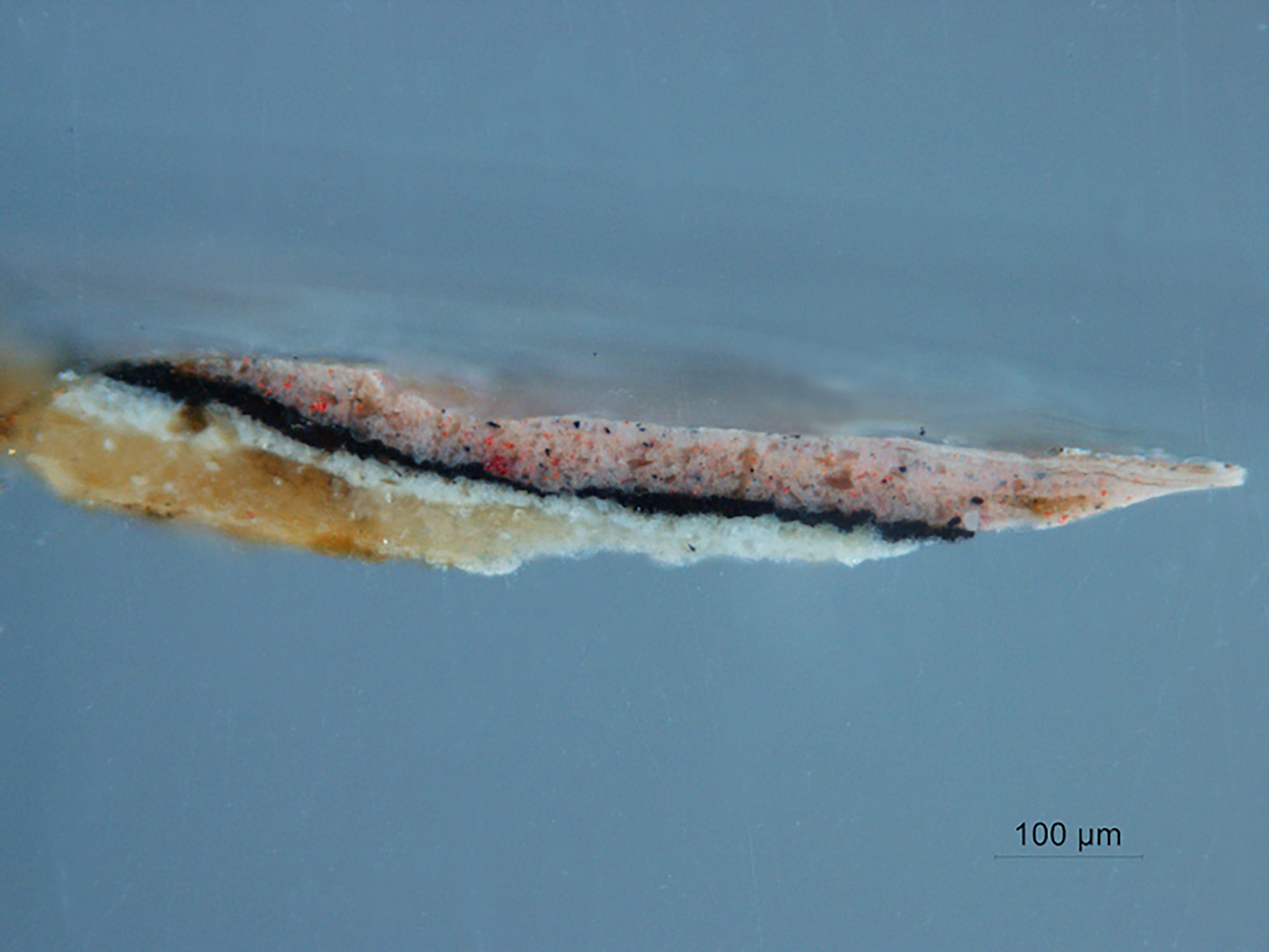
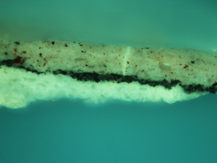
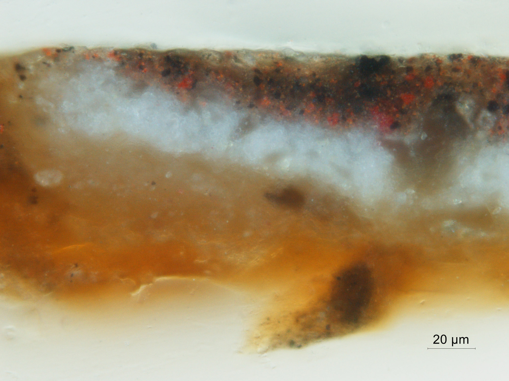
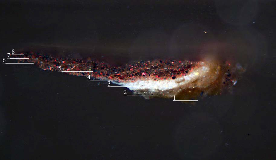
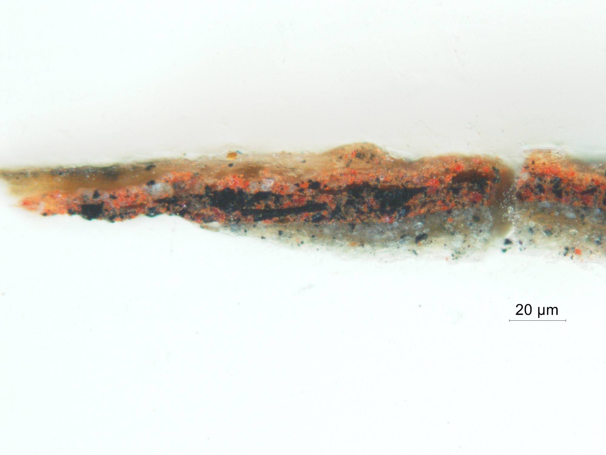
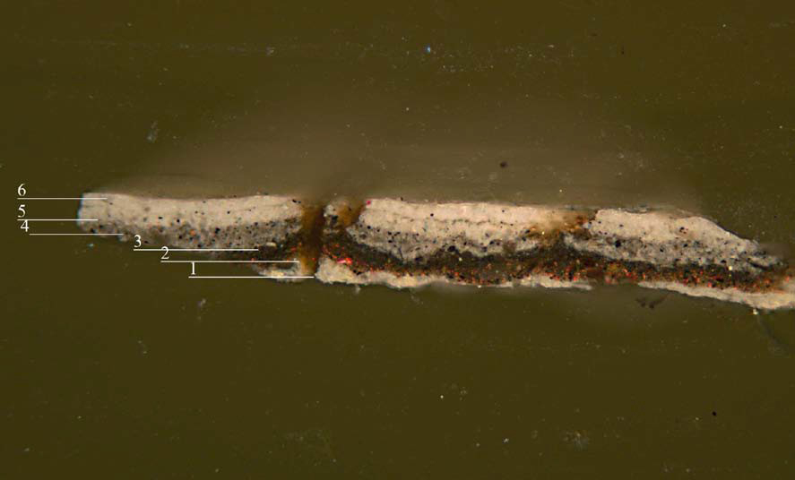
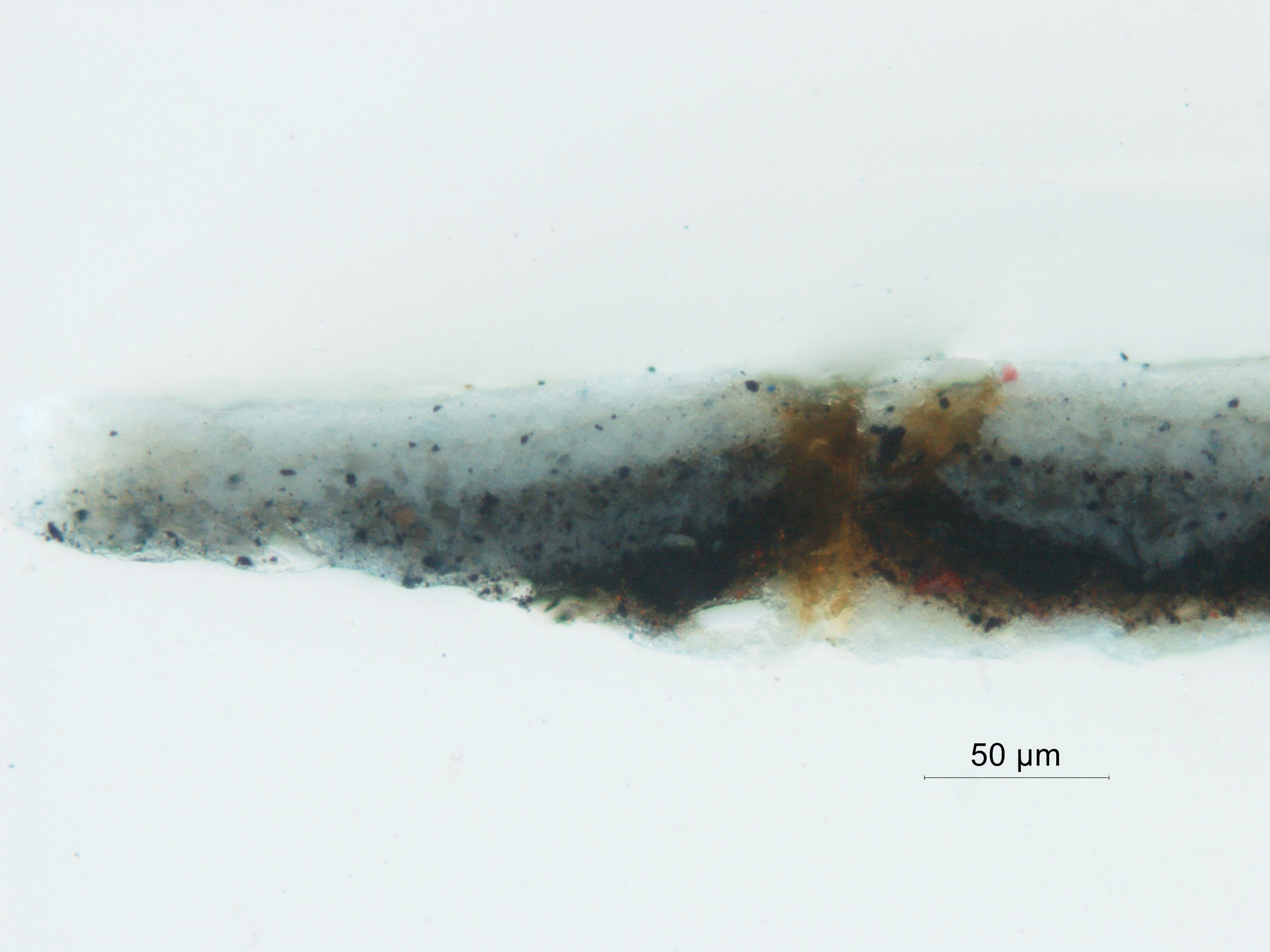
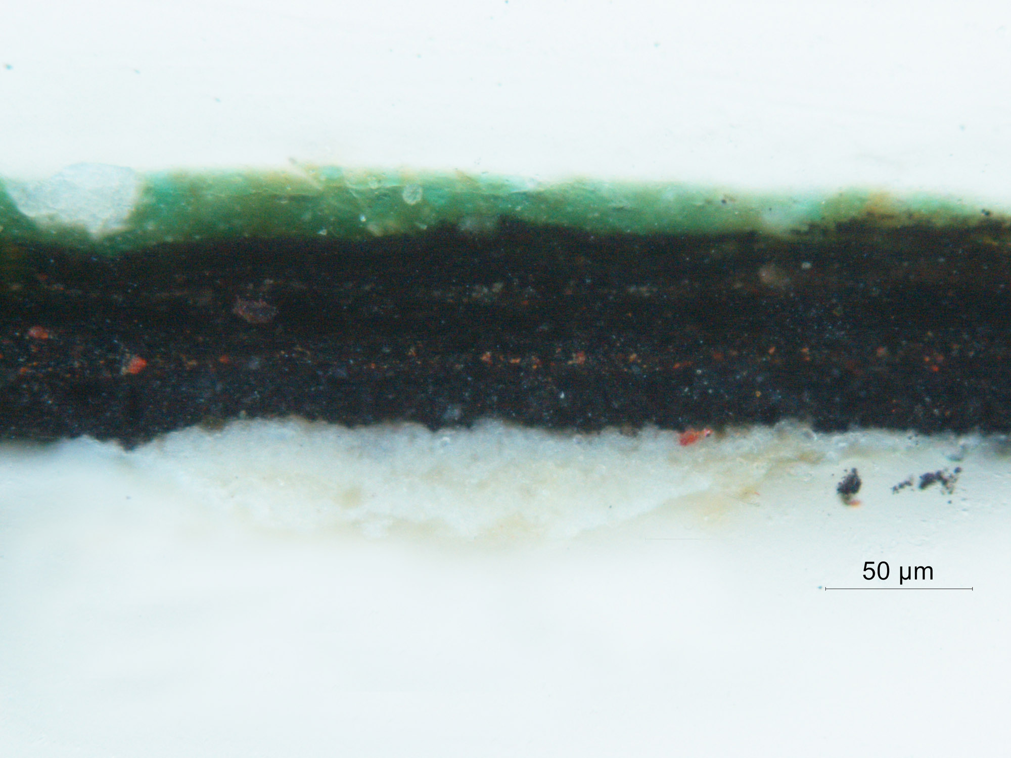
Sample 1, Highlight of Flesh Tone
-
Unpigmented layer with a slight orange fluorescence. Analysis with ATR-FTIR indicated a proteinaceous material, probably an animal glue size.
-
White preparatory layer containing lead white particles with soda lime glass containing manganese. Slight fluorescence under UV light.
-
Second preparatory layer containing finer particles of lead white and less soda-lime glass, the medium fluoresces brightly.
-
Black layer comprised of fine, densely packed black pigment particles. The paint layer is very lean and exhibits no fluorescence. SEM analysis confirmed the presence of bone black.
-
Thin light gray paint layer containing lead white and black pigment particles. The layer fluoresces under UV light
-
Warm, bright pink paint layer containing white lead particles, black, large red pigment particles of vermilion and glass. Fluoresces under UV light.
-
A slightly cooler layer containing the same pigments in slightly different proportions and tiny glass particles.
- Cool pink paint upper layers: 2 or 3 very thin layers (4-5 microns thick) visible at far right of sample. They contain fine particles of lead white, black and vermilion.
 Sample 1 annotated
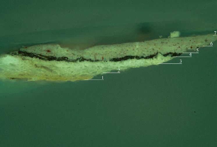 Sample 1 annotated, UV
 Sample 1, 10x
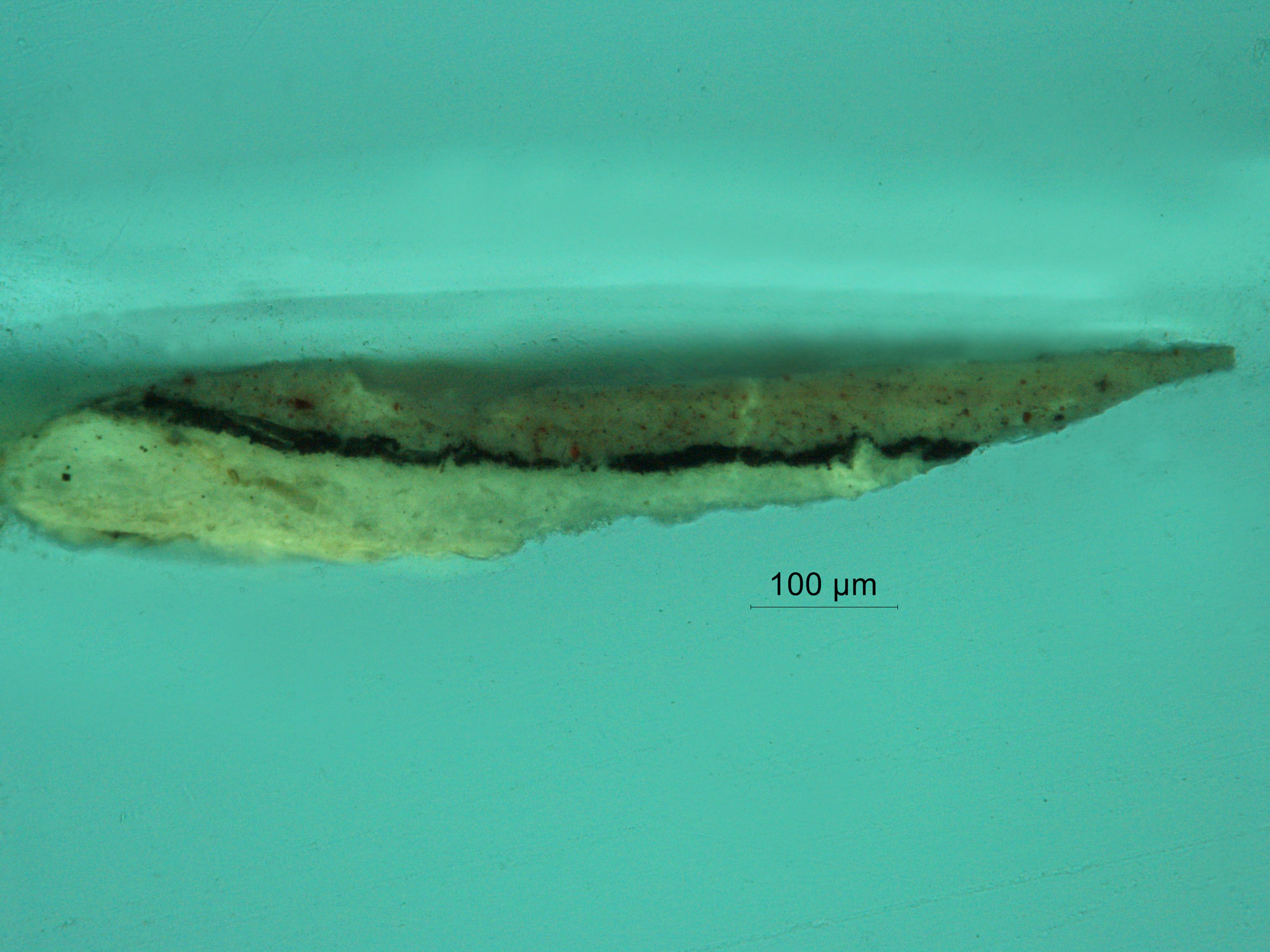 Sample 1, 10x, UV
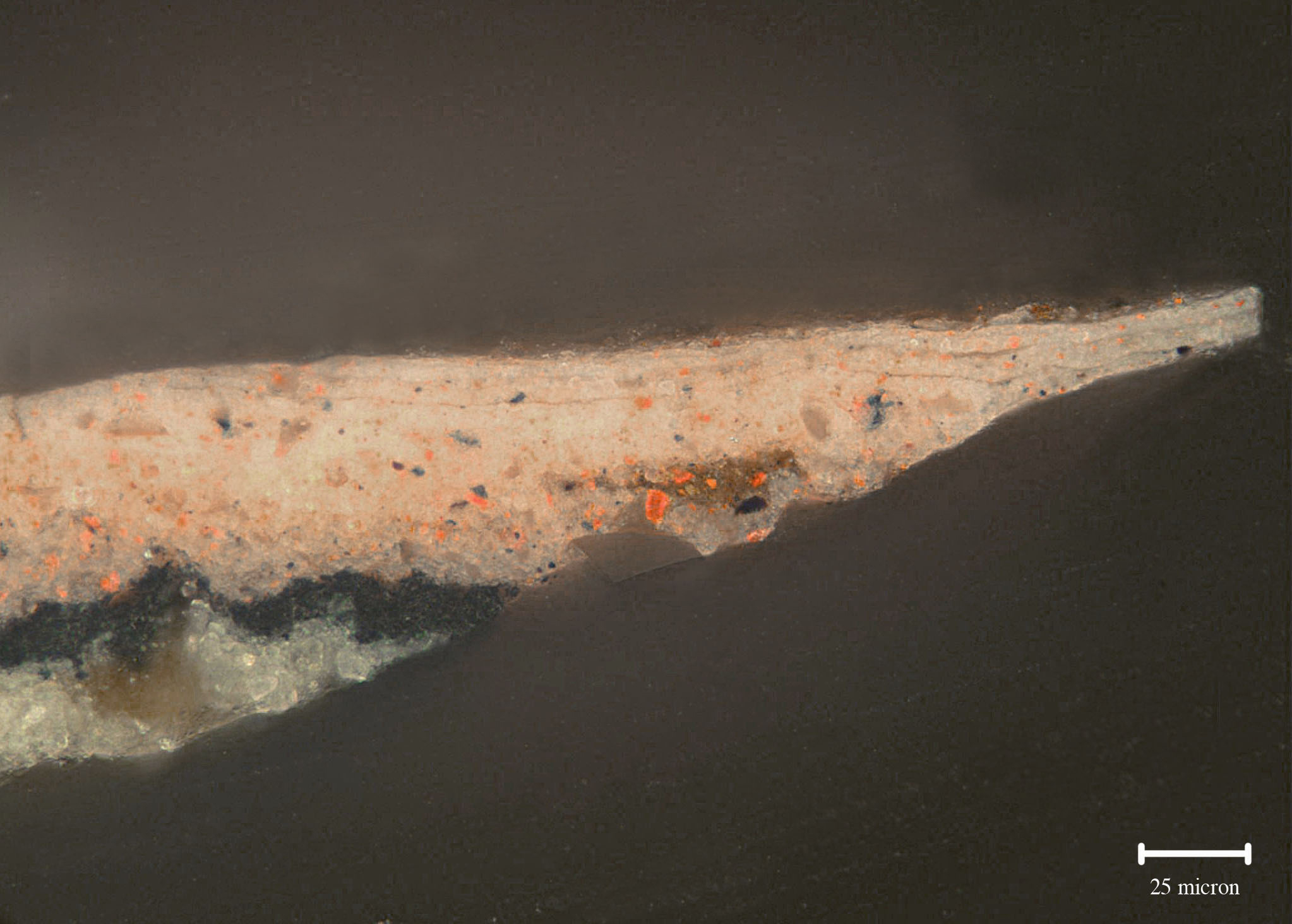 Sample 1, 4ox
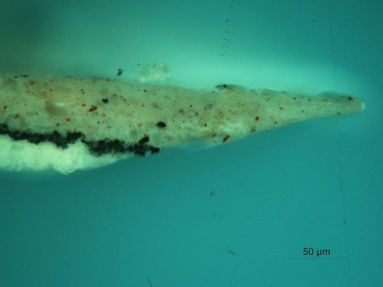 Sample 1, 40x,
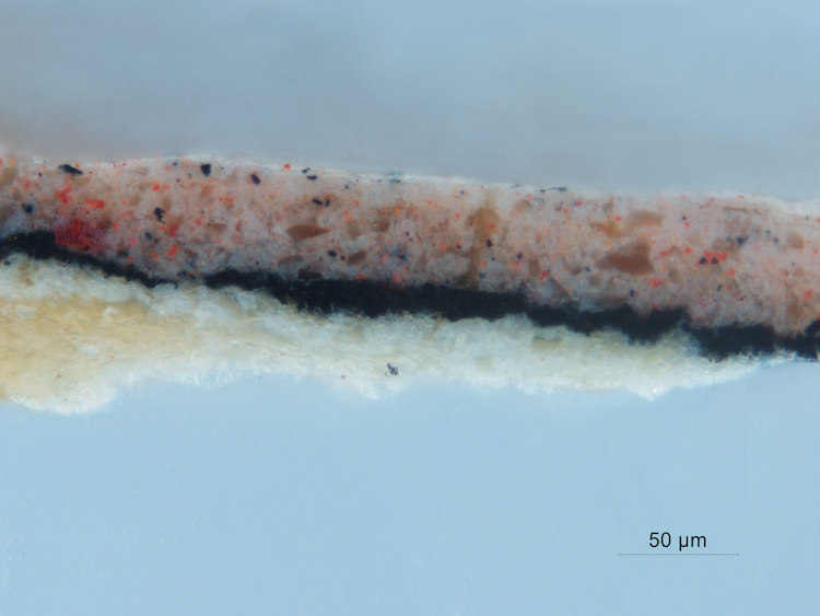
sample 1, 40X
 sample1, 40x, UV
Sample 2, Flesh Tone Shadow
-
Layer with a slight organge fluorescence, probaby an animal glue size.
-
White preparatory layer containing lead white and angular particles of soda -lime glass. Fluoresces brightly under UV light.
-
White preparatory layer containing finer particles of lead white and soda-lime glass. Fluoresces brightly under UV light.
-
Medium rich layer containing particles of vermilion and carbon black with pink fluorescence.
-
Another layer of vermilion and carbon black with a higher pigment/medium ratio.
-
Vermilion and a small amount of black with an overall pink fluorescence in UV light.
- Layer containing black and vermilion with a yellow/green fluorescence and similar to layer 5.
 Sample 2 annotated
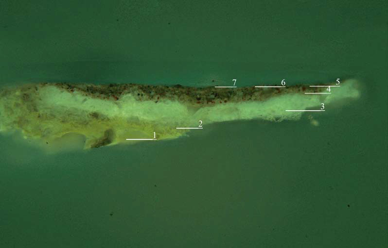 Sample 2 annotated, UV
 Sample 2, 25x
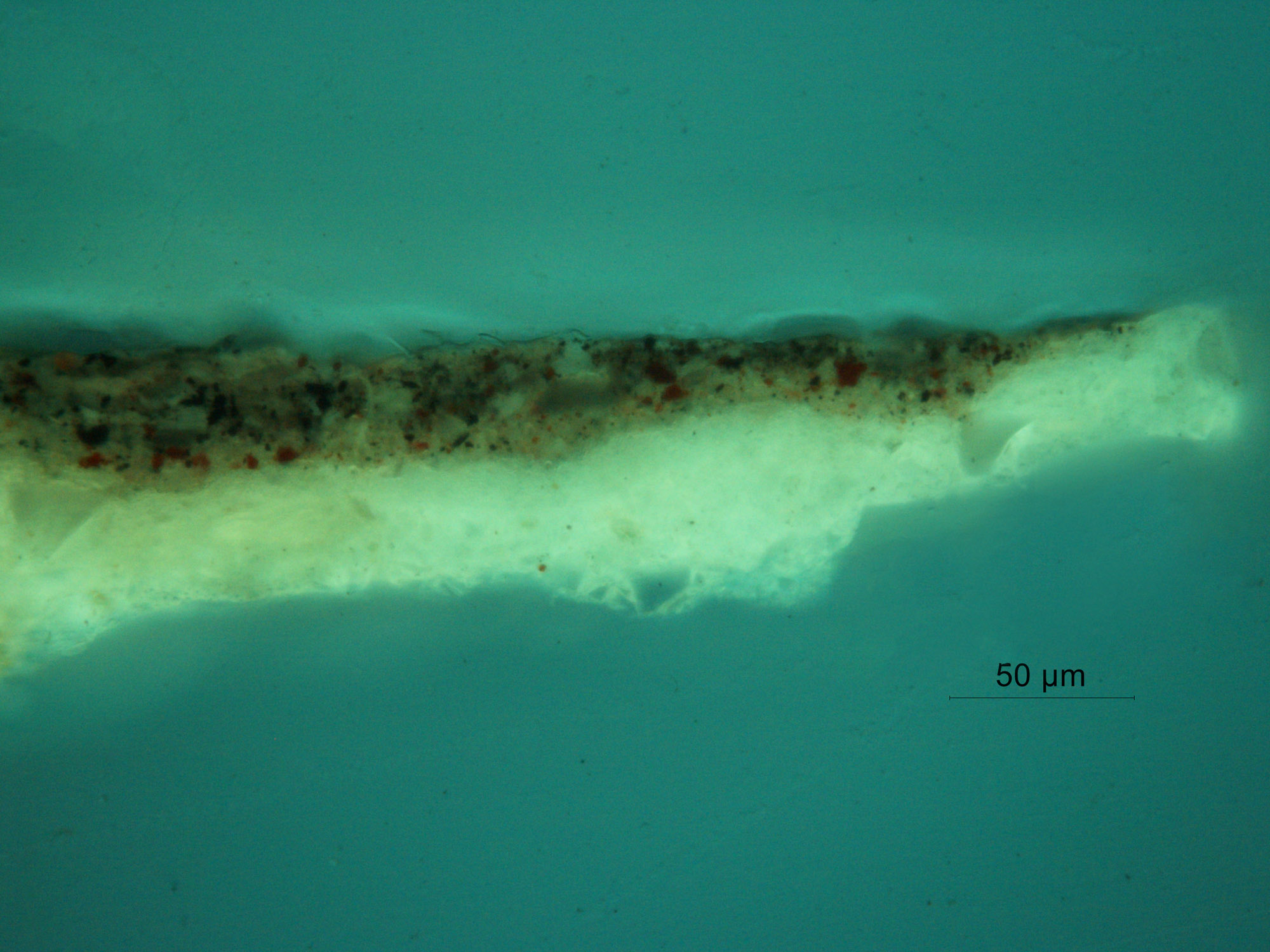 Sample 2, 25x, UV
 Sample 2, 40x
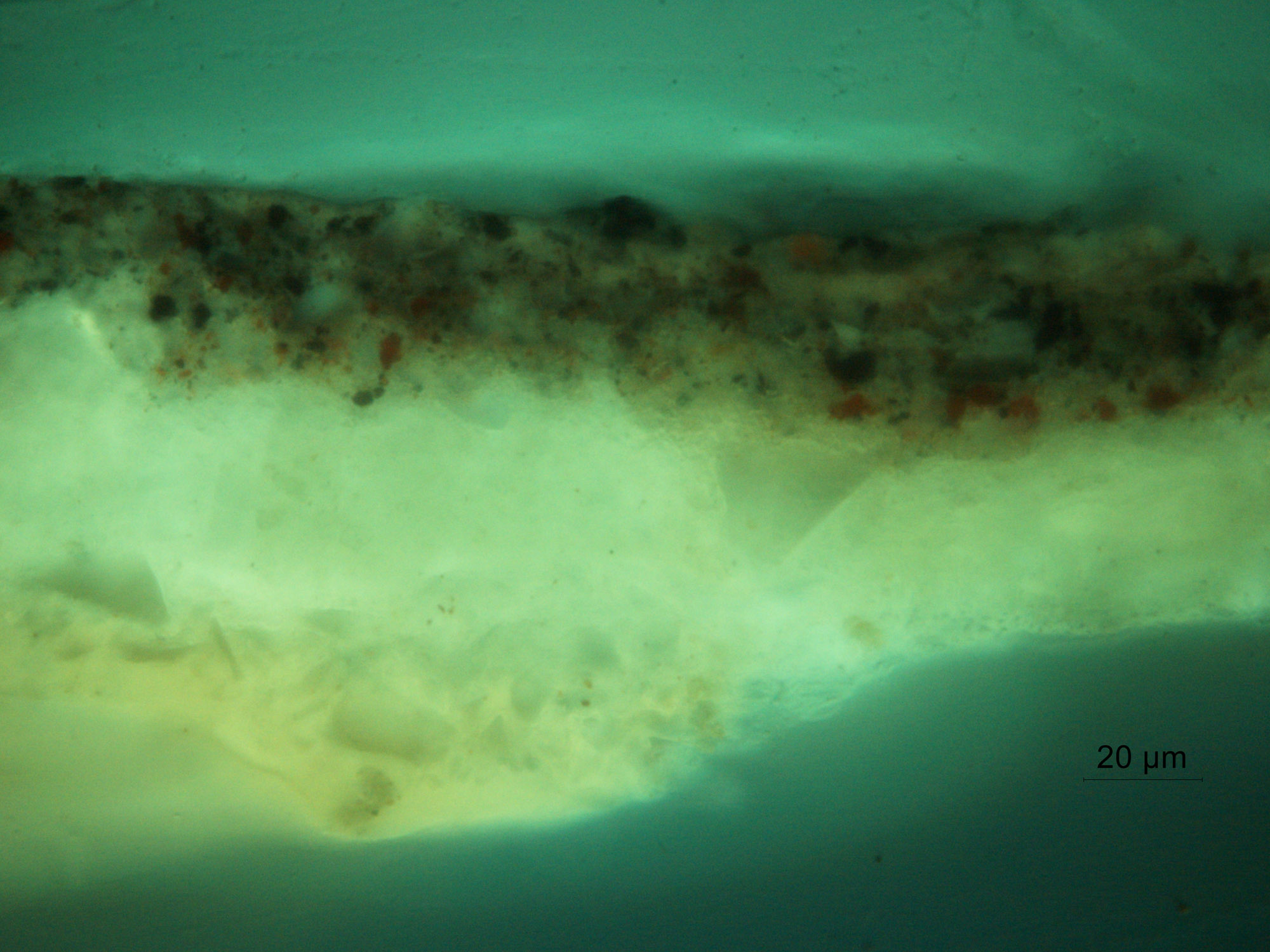 Sample 2, 4ox, UV
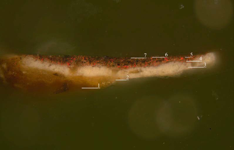
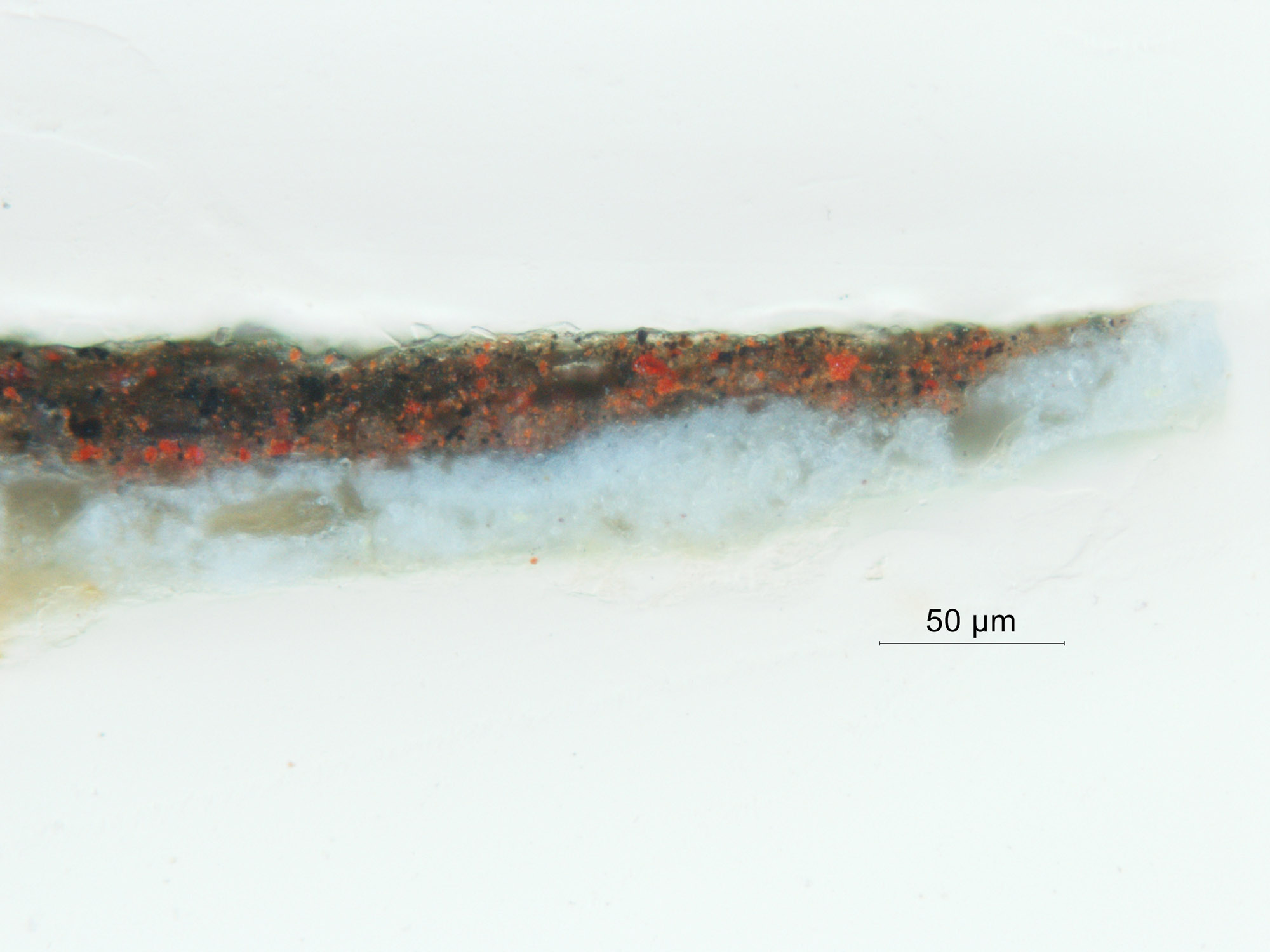
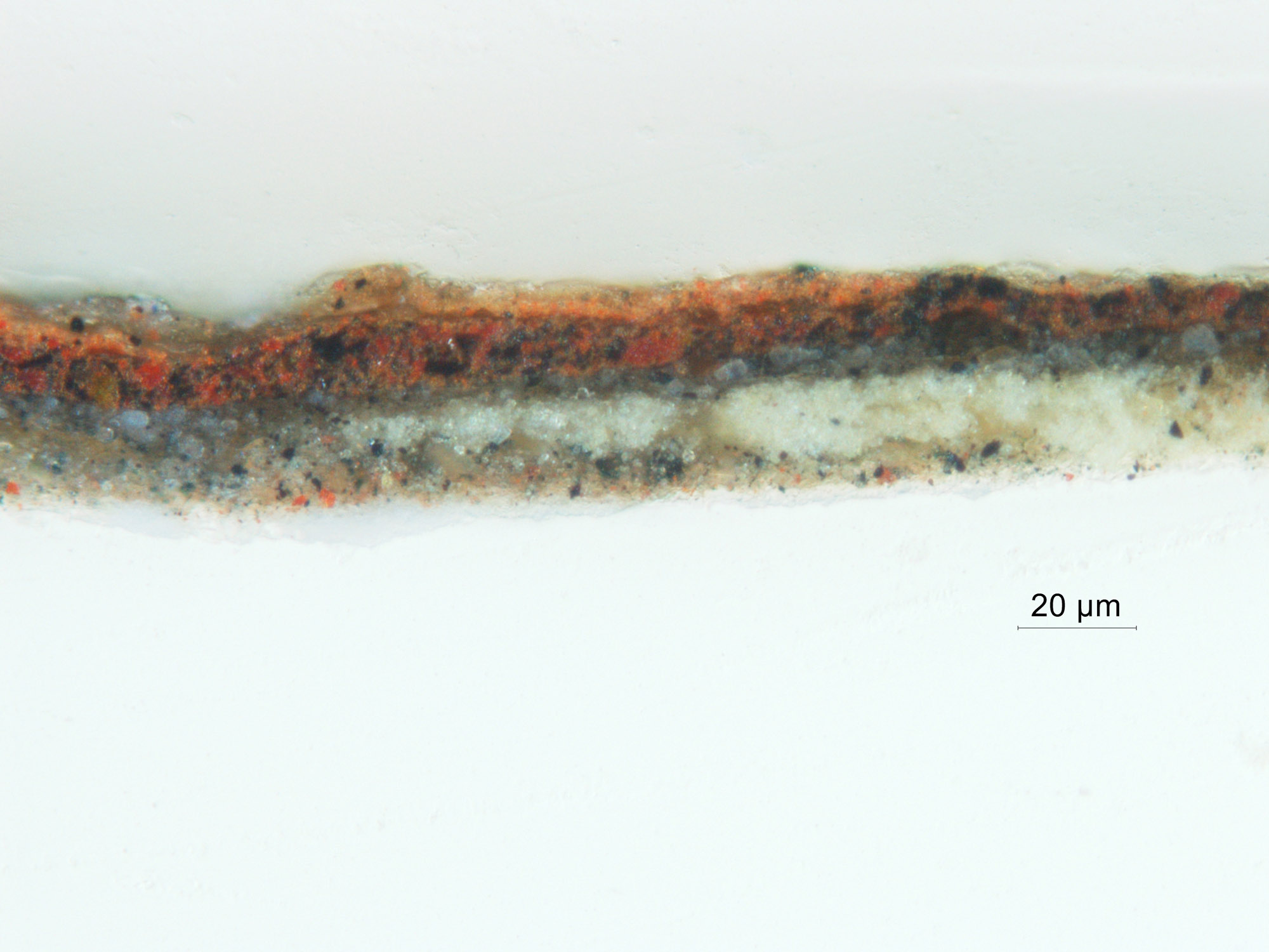
Sample 3, Flesh Tone Shadow 2
-
Unpigmented layer with a slight orange fluorescence, probably an animal glue size.
-
White preparatory layer containing lead white and angular particles of soda -lime glass. Fluoresces brightly under UV light.
-
White preparatory layer containing finer particles of lead white and soda-lime glass. Fluoresces brightly under UV light.
-
Medium rich layer containing particles of vermilion and carbon black with pink fluorescence.
-
Another layer of vermilion and carbon black with a higher pigment/medium ratio and pockets of a deep brown translucent layer that may contain a few particles of glass, no pigment and a strong yellowish green fluorescence.
-
Very thin layer containing just a few particles of vermilion and black, only distinguishable in UV fluorescence. Pink fluorescence.
-
Darker, reddish brown layer, similar to layer 5 with pockets of unpigmented medium.
- Thin warm brown layer with tiny black pigment particles and a strong pink fluorescence.
 Sample 3, annotated
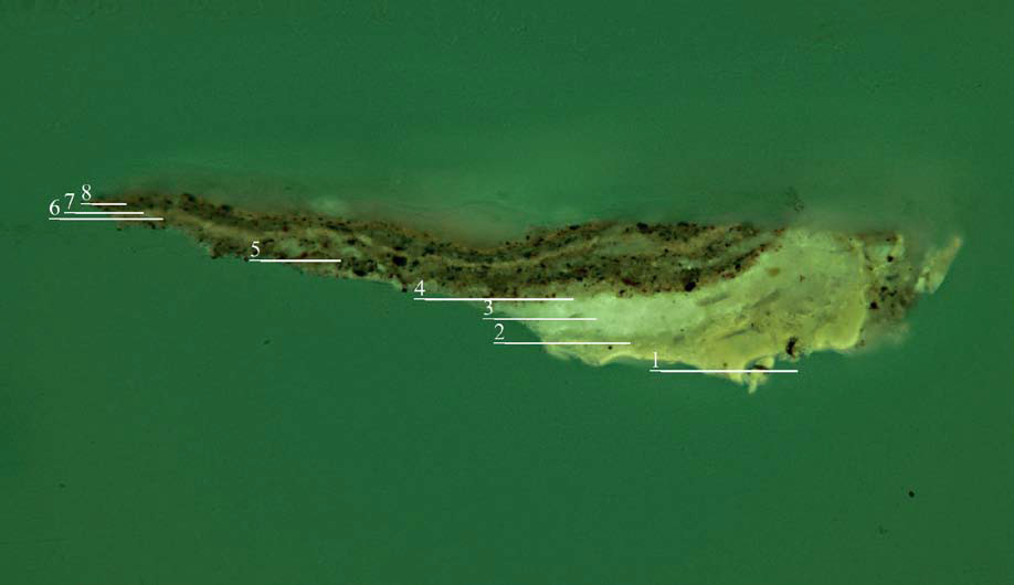 Sample 3 annotated, UV
 Sample 3, annotated, 4ox
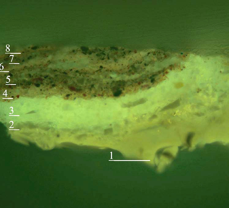
Sample 3, 4o annotated, UV
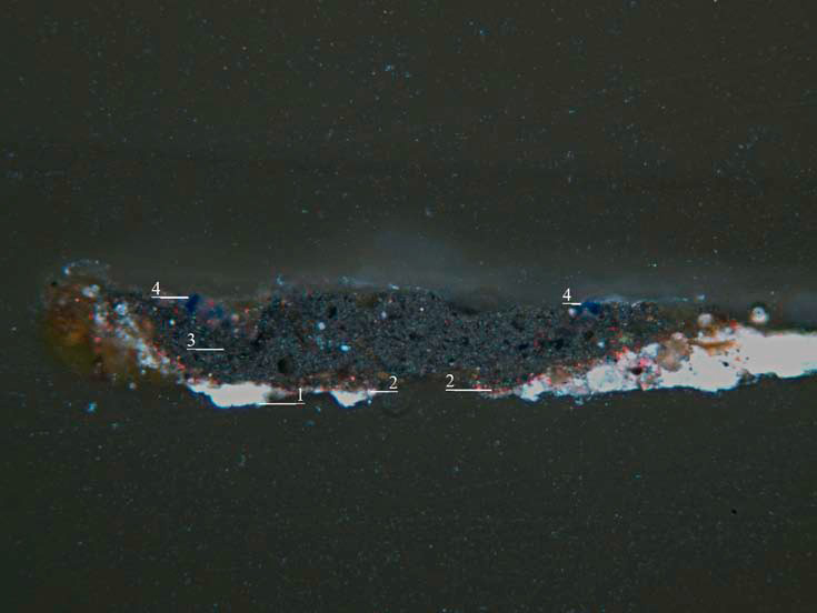
Sample 4, Blue Robe
- White preparatory layer of lead white and angular particles of soda -lime glass. Fluoresces brightly under UV light.
-
White preparatory layer containing finer particles of lead white and soda-lime glass. Fluoresces brightly under UV light.
-
A layer comprised of carbon black, vermilion and glass. Fluoresces under UV light.
-
Thin, uneven dark paint layer containing densely packed, tiny pigment particles in a mixture similar to the gray paint seen in a sample taken from the white sleeve and another from the barbe. Fluoresces under UV light.
-
Lapis lazuli pigment with particles of quartz. The layer has a slight pink fluorescence indicating the possible addition of red lake.
 Sample 4, annotated
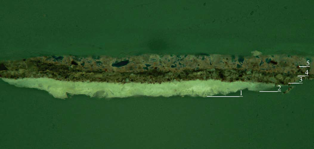 sample 4, annotated, UV
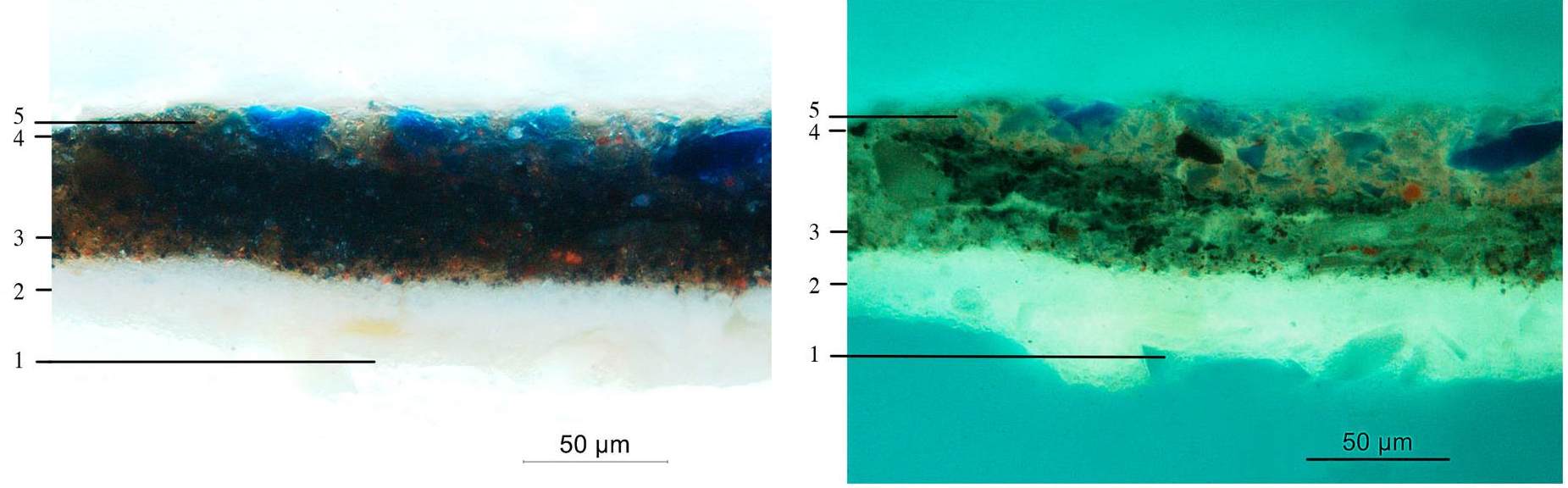 Sample 4, 25x, annotated
Sample 4, 4o x
 sample 4, 4o x UV
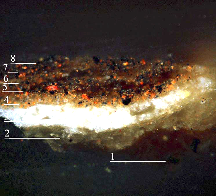
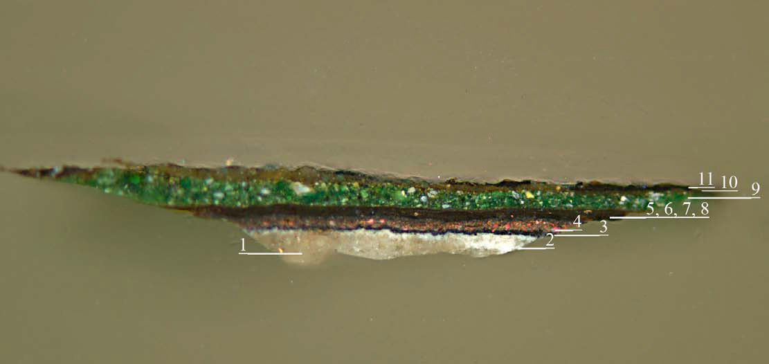
Sample 5, Knotwork
-
White paint layer. Lead white pigment particles very finely dispersed in fluorescent medium. Probably the upper ground.
-
Warm gray paint layer. Lead white pigments with tiny black pigment particles and small amount of vermillion. Slight fluorescence under UV light.
-
Pale yellow paint (first knot work).Lead tin yellow particles in oil medium with bright fluorescence in UV light.
-
Grey paint layer. Lead white pigment with tiny black pigment particles. Fluoresces under UV light.
-
Reddish brown paint layer. Densely packed vermilion, black and yellow pigment particles with very little medium. There is no fluorescence under UV light, which suggests that the binder might be proteinaceous.
-
Extremely thin, discontinuous unpigmented layer that fluoresces under UV light.
-
Reddish paint layer. Primarily vermilion with some scattered black pigment particles. Slight fluorescence under UV light.
-
Discontinuous unpigmented layer of substantial thickness on the far left and right ends of the sample with strong yellow-green fluorescence. Perhaps pockets of medium.
- A few remnants of a final paint layer, similar to layer 5. The sample was taken from an area of abrasion so little of this layer is left.
 Sample 5 annotated
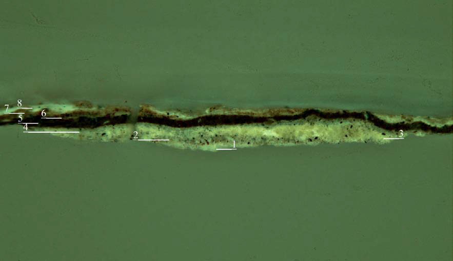 Sample 5 annotated, UV
 Sample 5, 25x
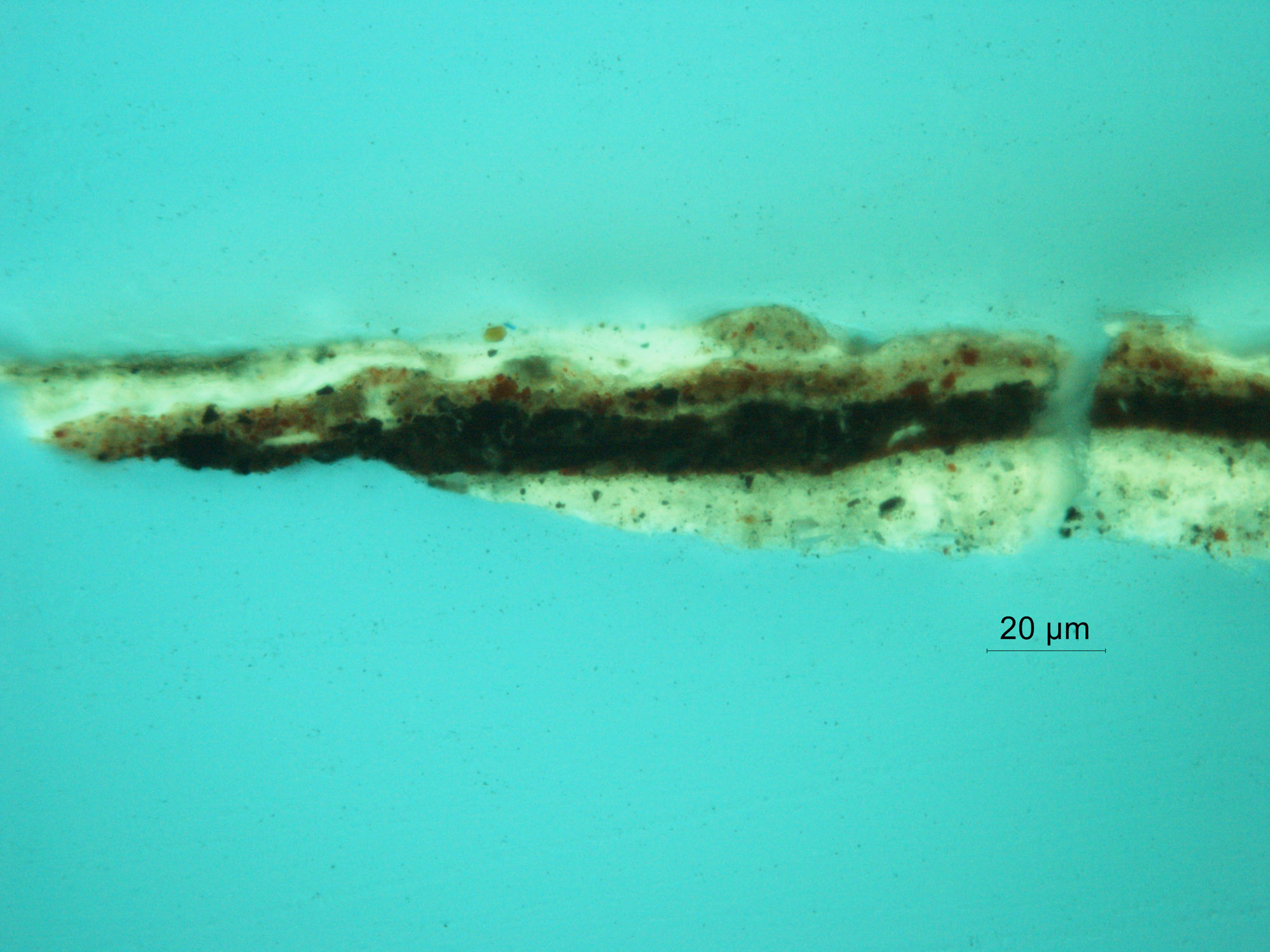 Sample 5, 25x, UV
 Sample 5, 25x
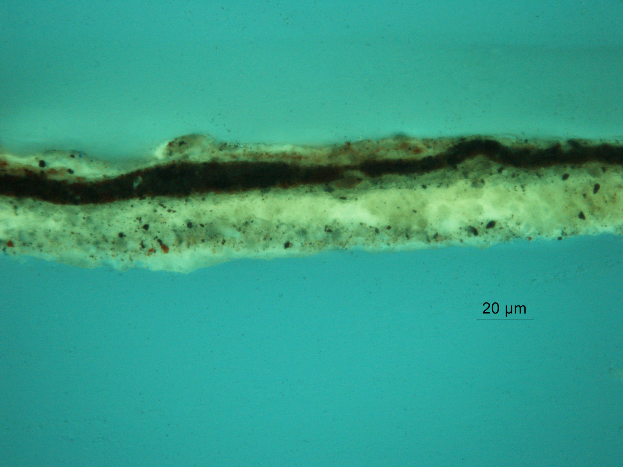 Sample 5, 25x, UV
Sample 6, White Sleeve
-
Preparatory layer containing lead white and particles of glass with bright fluorescence. Probably the upper ground.
-
Reddish brown layer with vermilion and black particles. The medium fluoresces yellow/green.
-
Black layer comprised of fine, densely packed black pigment particles. The paint layer is very lean and exhibits no fluorescence. SEM analysis confirmed the presence of bone black. This corresponds to the black wash of the initial
abozzo layer seen in Samples 1, 7, and 8.
-
Gray paint layer containing lead white and black pigment particles.
-
Light gray paint containing lead white and tiny black pigment particles. Fluoresces brightly under UV light.
- White lead paint layer with negligible fine scattered black pigment particles. Fluoresces brightly.
 Sample 6, annotated
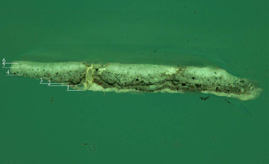 Sample 6, annotated, UV
 Sample 6, 25x
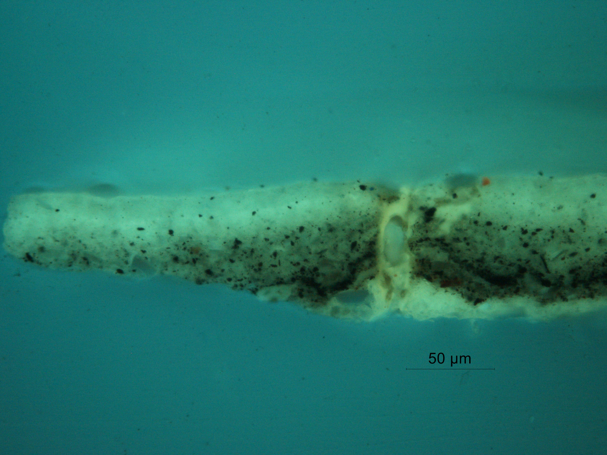 Sample 6, 25x, UV
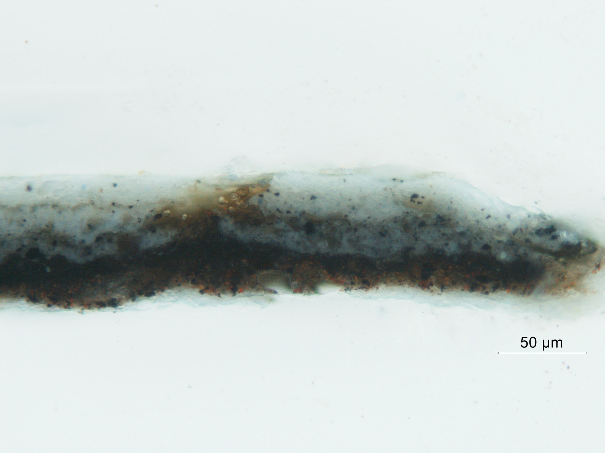 Sample 6, 25x
 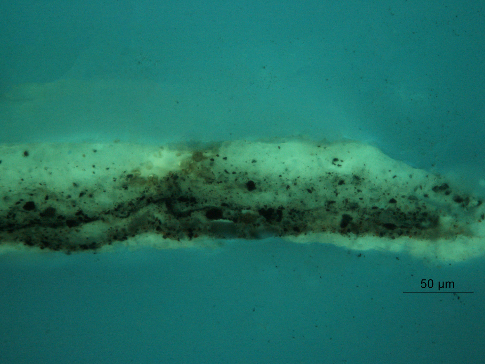 Sample 6, 25x, UV
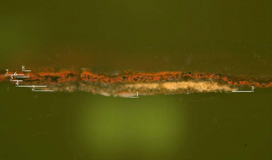
Sample 7, Background Under Green Repaint 1
-
White preparatory layer containing lead white and angular particles of soda -lime glass. Fluoresces brightly under UV light.
-
Reddish brown layer with vermilion and black particles. The medium fluoresces yellow/green.
-
Second white preparatory layer containing finer particles of lead white and less soda-lime glass. Fluoresces brightly under UV light.
-
Reddish brown layer composed of carbon black, vermilion and possibly a lake pigment (suggested by the pink fluorescence under UV light.)
-
A thin layer of tiny black pigment particles with no fluorescence.
-
Mixtures of carbon and bone black with some fluorescence.
-
Thin layer of black pigment.
-
Unpigmented fluorescent layer.
- Green layer containing verdigris, yellow and a few particles of white.
 Sample 7 annotated
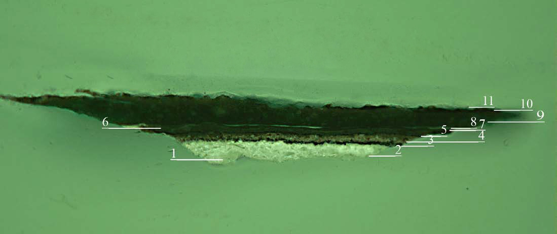
Sample 7 annotated, UV
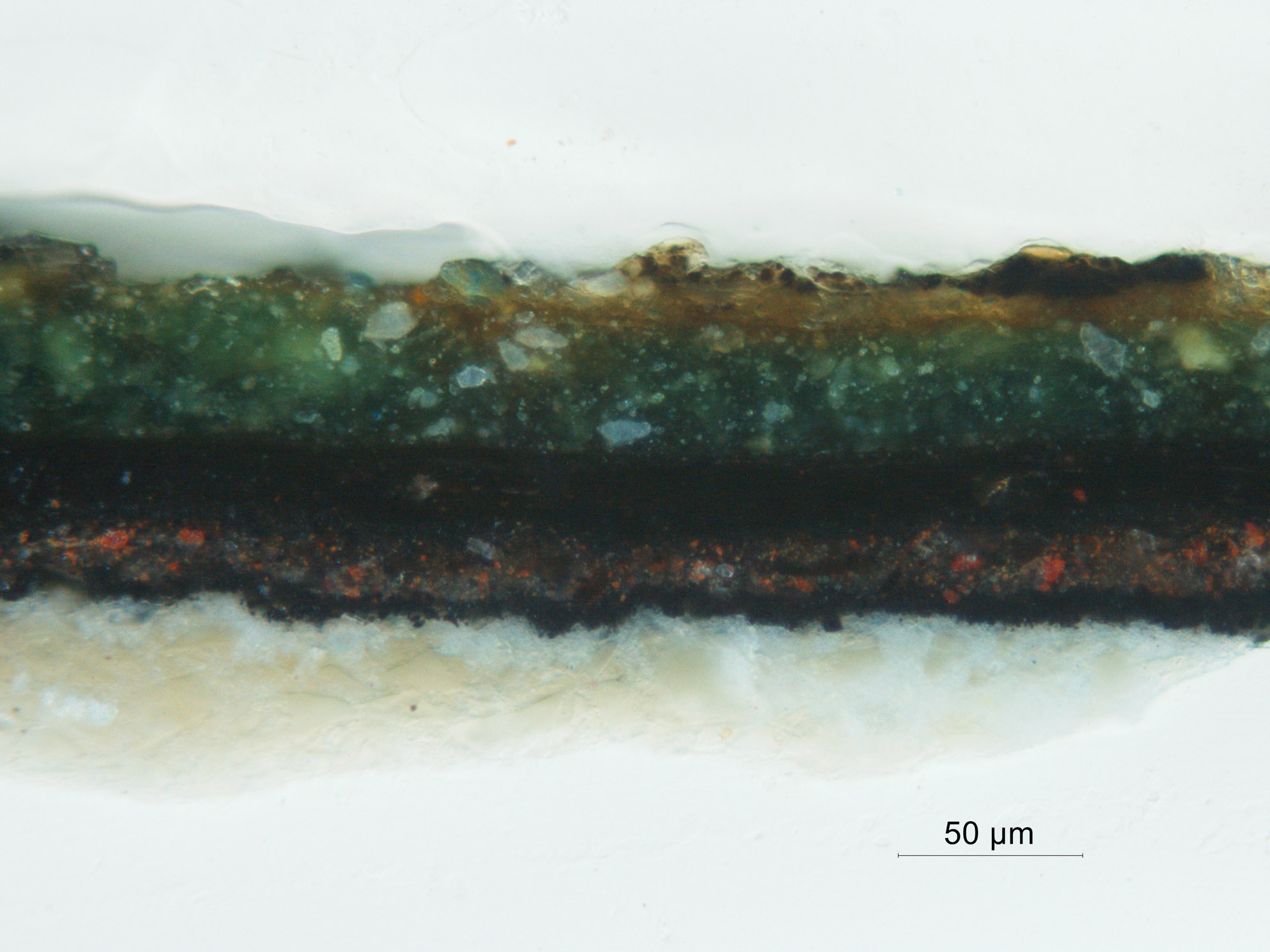 Sample 7, 40x
 Sample 7, 40x, UV
 Sample 7, 25x
 Sample 7, 25x, UV
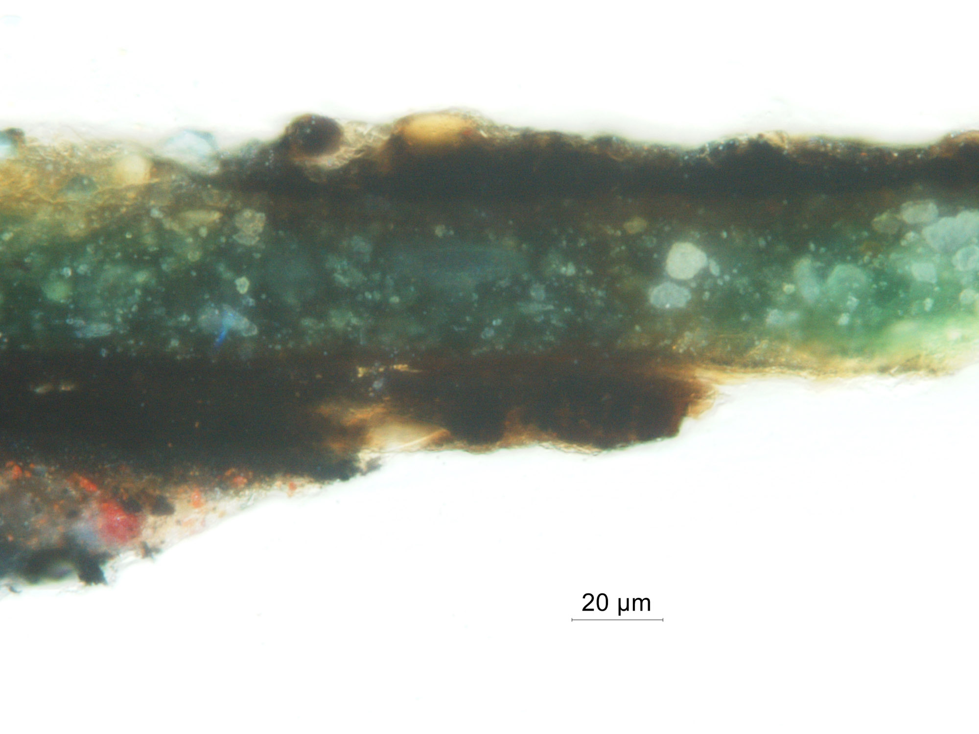
Sample 8, Background Under Green Repaint 2
-
White preparatory layer with lead white and angular particles of soda-lime glass. Bright fluorescence under UV.
-
Second white preparatory layer Upper ground containing more finely ground particles of lead white and soda lime glass. Fluoresces brightly under UV light.
-
Black layer comprised of fine, densely packed black pigment particles. The paint layer is very lean and exhibits no fluorescence.
-
Reddish brown layer containing vermilion and black pigment particles. Fluoresces under UV.
-
A thin layer of tiny black pigment particles with no fluorescence.
-
Mixtures of carbon and bone black with some fluorescence.
-
Thin layer of black pigment particles with no fluorescence.
-
Unpigmented layer with bright yellow/green fluorescence under UV light.
-
Green paint layer. Thick layer composed of green (verdigris), yellow and white pigment particles. There is a remnant of dark material on top of the left side of the sample.
-
Thin layer with slight fluorescence and no particles.
- Dark, discontinuous paint layer with no fluorescence.
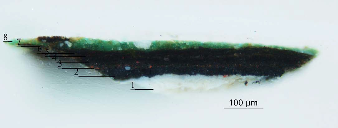 Sample 8, annotated
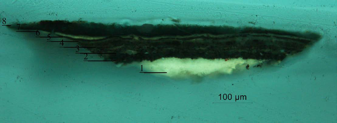 Sample 8 annotated, UV
 Sample 8, 4ox
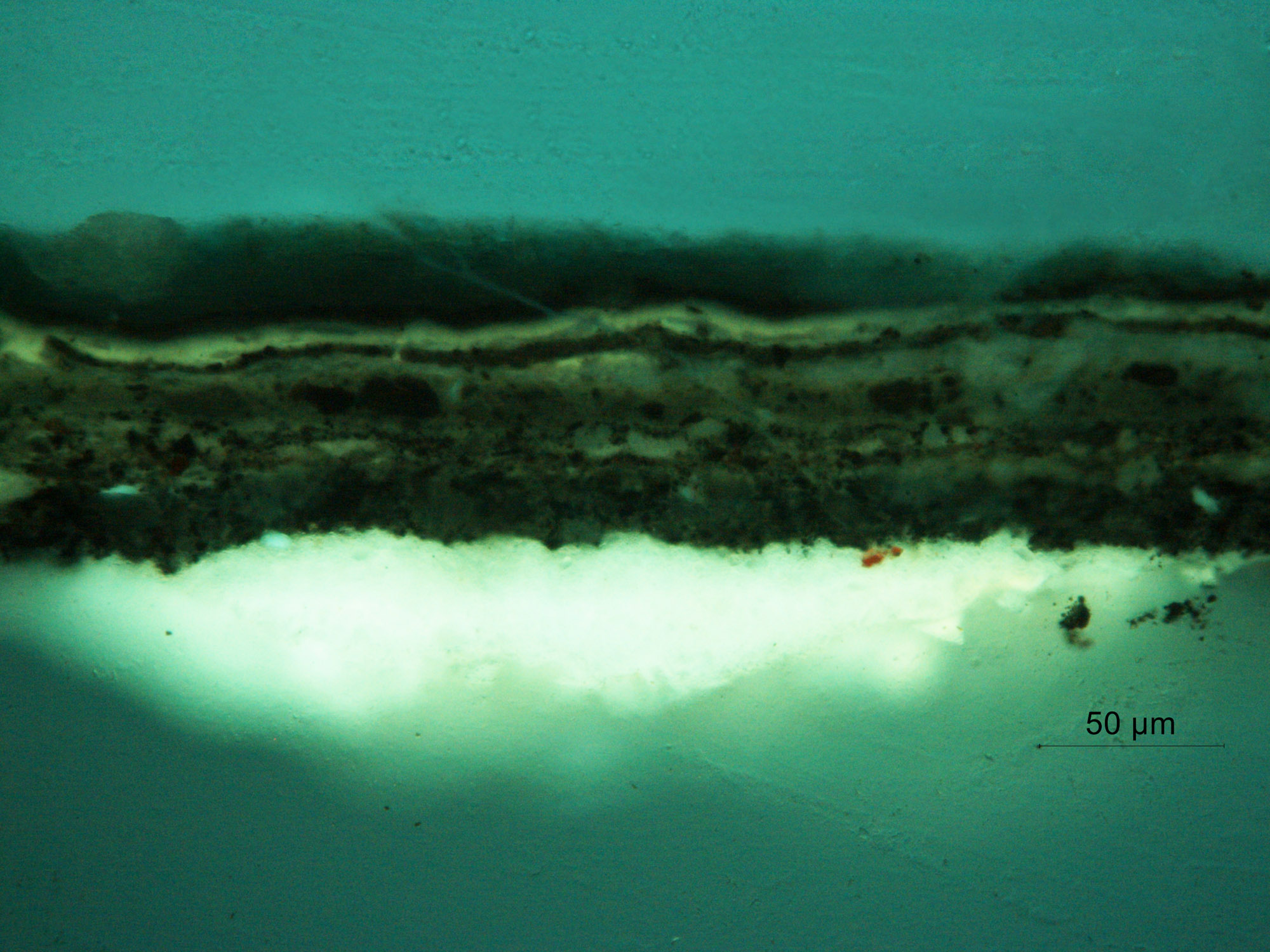 Sample 8 40x, UV
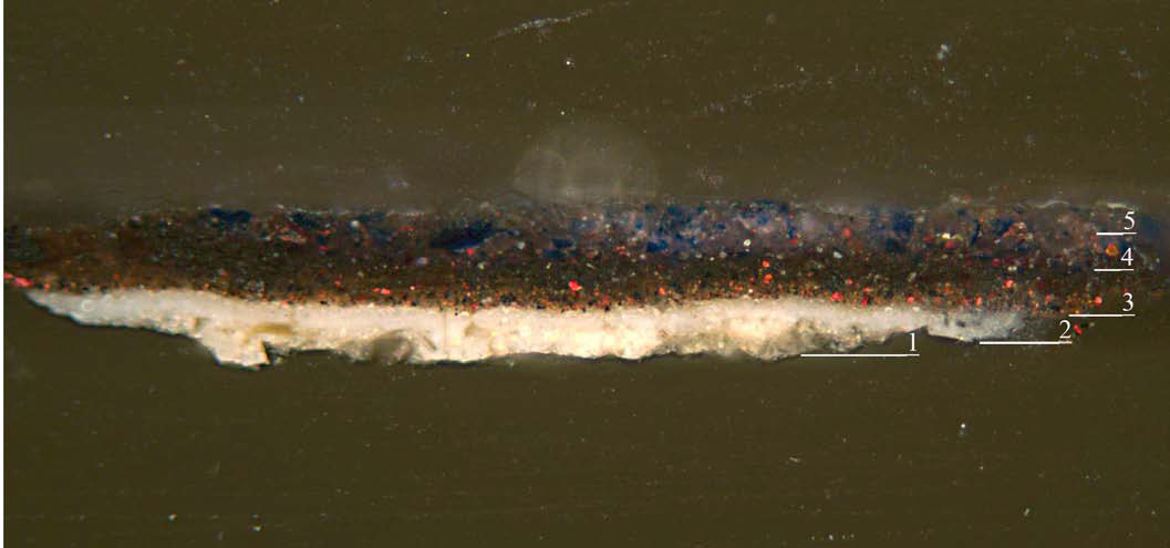
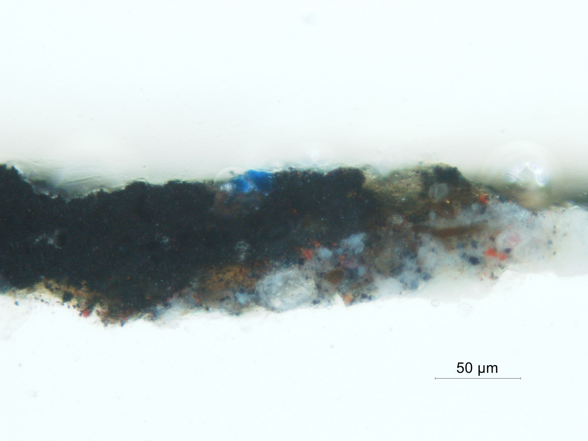
Sample 9, Barbe, Bottom Left Edge
-
White preparatory layer containing lead white and soda lime glass. Fluoresces brightly under UV light.
-
Reddish brown paint layer containing a high proportion of medium with some suspended particles of vermilion and black.
-
Black layer comprised of fine, densely packed black pigment particles. The paint layer is very lean and exhibits no fluorescence.
-
Gray paint layer, very thick as it is an accumulation along the barbe. It is marbleized with medium with bright yellow/green fluorescence under UV light.
- Blue paint with large particles of lapis lazuli in a medium that fluoresces yellow green. There are also some particles with strong pink fluorescence characteristic of a red lake.
 Sample 9 annotated
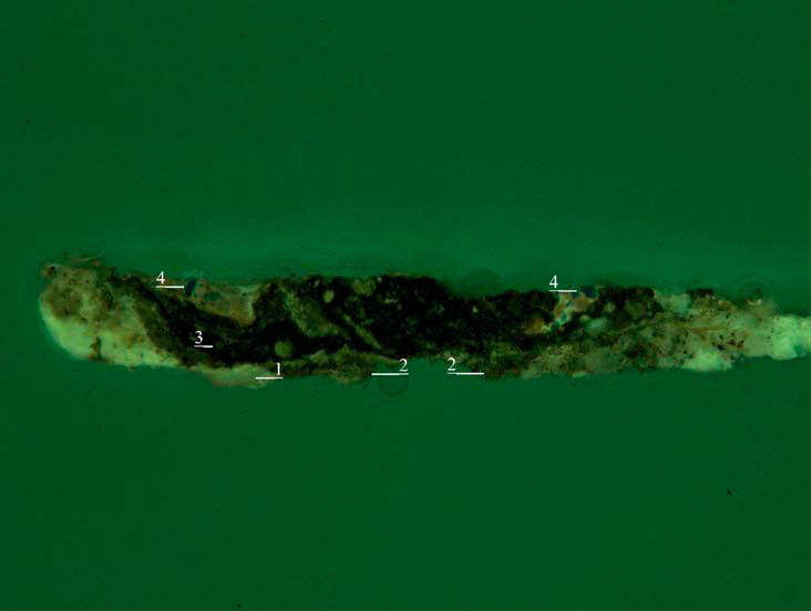 Sample 9 annotated, UV
 Sample 9, 25x
 Sample 1, 25x, UV
Media Analysis
Walnut oil was identified in a sample taken from the lower right quadrant of the painting near the Py-GCMS analysis was carried out at the Philadelphia Museum of Art as follows: the sample (c. 10 ug) was placed in a Frontier Lab stainless steel sample cup, and 2uL of a 25% solution of tetramethylammonium hydroxide (TMAH) in methanol added prior to insertion into a Frontier PY-2020iD vertical micro furnace pyrolyzer, attached to an Agilent 6890N GC - 5973N MS. The pyrolyzer furnace temperature was 550°C. The GC was equipped with J&W DB5MS column (30 m, 0.25 mm i.d., 0.25 um film); oven temperature programmed from 40°C, with a 2 minute hold, then increased at 20°C/minute to 140°C, at 15°C/minute to 320°C, and held isothermally for 11 minutes; total run time 30 minutes. The inlet was operated with a split ratio of 1:10, with helium carrier gas at a flow rate of 1 mL/minute. The MS was run in scan mode (m/z 35-600) with the source at 230°C and quad at 150°C. Data were collected and processed using Agilent Chemstation software.